2011年4月17日 星期日

Proteinuria


  1. 正常尿中蛋白約45~150mg/天,運動後會到300。其中有三成是albumin,其他為globulin。
    • 一般做dipstick的結果表示:trace(10mg/dl),+1(30mg/dl),+2(100mg/dl),+3(300mg/dl),+4(≧1000mg/dl)
    • 當light chain的globulin太多時,dipstick會低估實際值(dipstick主要檢查albumin)。
    • 7%女性有transient proteinuria,而男性為4%。
  2.  檢查:收24小時尿液最簡單,也要做24小時creatinine excretion。結果可分三種:overflow:plasma protein↑;tubular:tubular再吸收小分子量protein有問題;glomerular:只有glomerular常有>2g/天的結果,其他很少
  3. Multiple myeloma:屬於overflow型,有很多單股的 κ, λ light chain。
    • 10%MM有amyloidosis,會loss到3g/天。
    • dipstick對light chain沒反應→false negative
  4. β2-microglobulin:分子量12000,是測tubular的marker。正常約100μg/天。tubular的疾病會有10倍~100倍的增加
  5. hereditary tubular disease: PCKD, Wilson's disease, cystinosis, oxalosis, medullary cystic disease
  6. congnital tubular disease: 部分RTA,所有的Fanconi's syndrome
  7. acquired tubular disease: 重金屬,PN,interstitial nephritis, obstructive uropathy, radiation nephritis, Vit.D 中毒等等
  8. immunologic tubular disease: 腎臟移植排斥,sarcoidosis,藥物過敏,Sjogren's disease
  9. 感染:會有nephrotic-range proteinuria的疾病有:PSGN,IE,shunt nephritis syphilis,病毒(HIV, HBV, HCV)
  10. 藥物:會超過3g/天。有可能是penicillamine, NSAID, mesantoin, probenecid
  11. 家族性的:Alport's syndrome, sickle cell disease, Fabry's disease, nail-patella syndrome
  12. 癌症:
    • 直接毒害kidney的有lymphoma, leukemia, Wilm's tumor, pheochromocytoma, MM
    • 間接造成glomerular damage:membranous nephropathy,通常跟lung, breast, colon的癌症有關
  13. GN:
    • 致病機轉至少有兩種:血管壁的副電荷消失。大分子的barrier改變
    • 可能是上皮細胞剝落,BM物質的沉積造成glomerular basement membrane gap的改變
  14. 會影響多個系統的疾病:如腎血管外傷,抗體沉積在腎絲球,或是抗原抗體複合物沉積在腎絲球等等,機轉同上。

Oliguria

  1. Oliguria:每天尿量<400ml,常是GFR↓所造成。正常血中溶質約400~500mOsm/天,尿最多壓縮到1200mOsm/kg,尿<400ml/天則無法排出全身的鈉。同時含氮物也會累積~BUN↑
  2. UNa可以反映出renal perfusion,只要tubular function正常的話,測FENa會更準。perfusion↓會刺激鈉再吸收。通常underline會有:腎血流↓(CO↓, 腎血管阻塞),管內volume depletion。
  3. prerenal的問題是最常見的原因。其中包括CO↓, diuretics使用過度,要治療underlying才行。會造成腎衰竭的藥物:prostaglandin synthesis inhibitor(propionic acid類的ibuprofen, naproxen;indoleacetic acid類的indomethacin),或是ACEI類的藥物。這些藥會↓GFR,腎血流。病人effective volume↓或是有腎血管疾病時會更糟糕。
  4. GN:glomerular inflammation會有nephritic urine sediment。可能有RBC, WBC, RBC cast, hyalin cast, cellular debris。除了RBC cast較有特異性,出現其他的東西可能是其他問題。
  5. 也常跟RPGN有關,病人常有自體免疫的問題,會有anti-GBM(+)或idiopathic crescentic GN。RPGN可能在streptococcus, HBV, IE後發生。其他相關的疾病有SLE, HSP, systemic necrotizing vasculitis, polyarteritis, cryoglobulinemia等
  6. 腎臟超音波可用,是一個noninvasive的工具。可看到calyces, pelvis擴張的話表示有extrarenal obstruction。但一開始24~36小時可能看不到,所以要重複測。
  7. 也可能是跟glomerular injury無關的vasculitis。如:idiopathic systemic vasculitis, Wegener's disease, granulomatosis, scleroderma vasculitis, hemolytic-uremia syndrome, 惡性高血壓,或是藥物。
  8. Gallium scan(+)表示有inflammation,可能是intterstitial nephritis或是infection。尿中可看到eosinophil偏向allergic。
  9. allergic form常跟藥物有關。interstitial nephritis跟antibiotics, NSAID, thiazide, allopurinol, aspirin, cimetidine, methyldopa, phenytoin, clofibrate等有關係。
  10. nonallergic form的原因多是infection。如streptococcus, toxoplasma, Legionnaire's, Rocky Mountain spot fever等
  11. acute tubular necrosis的ischemic form。常見原因:因volume loss導致的hypotension,CO↓,sepsis的vasodilation, rhabdomyolysis, postpartum hemorrhage, pancreatitis等等。prerenal的問題沒治療好也會變成ischemic form。
  12. toxin:常見的有aminoglycoside, amphotericin B, vancomycin, foscarnet等。也可能是重金屬(ex. Pb, Hg, cisplatin),endotoxin, myoglobin, Bence Jones protein, 含碘的contrast, fluorinated麻醉藥, 有機溶劑, ethylene glycol, Panadol, paraquat等

Hematuria

  1. haematuria:尿中太多RBC。正常一天最多10^6個。若有 >2~3RBC/HPF要進一步survey~收中段尿最準
  2. 引起haematuria的藥物:analgesics, cyclophosphamide, broad-spectrum penicillin, vincristine,口服避孕藥等等
  3. 若病人有在吃抗凝血藥,有凝血方面的疾病,就可能有自發性血尿。 但一些研究說這些人中過半數也要尿路系統的問題,所以應該排IVP,cystoscopy檢查。
  4. 有sickle cell的病人常有血尿,且HbAS比HbSS的更多。大多無痛,有80%來自左腎,同上,也很可能有尿路問題。
  5. 尿蛋白>1g/天表glomerulus受損。雖然有些腎絲球疾病尿protein不會高,但這些病也少有血尿
  6. 用更好的影像工具做診斷。ex. CT-enhanced excretory urogram, spiral CT
  7. 在腎臟tumor中RCC占了85%,60歲為高峰期,抽菸的人得到的機會是普通人的2~8倍。含nitrosamine的東西(ex. coffee, 高脂食物)也可能是致癌因子。
  8. 單獨renal cyst少有血尿,但10~15% PCKD會有(cyst內積血)
  9. 4%尿路tumor長在urothelial,TCC佔urothelial tumor的90%。剩下大多是SCC,adrenal carcinoma很少見。患者多60歲,男:女=4:1
  10. bladder tumor男人較多,吸菸也是危險因子。85%膀胱癌為TCC。
  11. acute/chronic GN都跟血尿有關。renal parenchymal來的血尿會有RBC cast, smoky。血尿不能看出是哪種疾病,但常見的有:MPGN, focal glomerular sclerosis, IgA nephritis (Berger's)

2011年4月16日 星期六

腹部X光基本判讀

  • 一張X光片大小為35x43公分,照腹部通常要兩張
  • KUB: Kidney -Ureter-Bladder。通常躺著做,bowel gas最好少些,較好看
    plain Abdomen: 看obstruction,常站立做,air-fluid level才好看
  • 氣體(黑色),fat/soft tissue/fluid(淺灰),骨頭/鈣化(白色),金屬(純白)
  • 女生要確定沒有懷孕。若是trauma,loss of consciousness的病人做尿液分析排除
  • 左右分別:
    左:腎臟較高,胃,脾,心臟
    右:肝臟
  • 基本上要看的東西-實質器官
  1. 看骨頭
    • 由骨盆看性別
    • osteoporosis
    • sarco-ilitis:多與腸道疾病有關。ex. Crohn's disease,膽結石發生率↑
    • Paget's disease, myeloma, metastatsis
    • 注意骨頭上的腸氣可能會干擾蝕骨性lesion的判斷
  2. 看iliopsoas:
    Silhouette sign輪廓徵象。通常iliopsoas旁邊為fat圍繞,X光片中可看到邊緣,但如果有出血,膿,腫瘤的話,psoas就會看不清楚。但也可能是良性的,如gas太多,脊椎側彎,fat太少等。
  3. 看kidney:
    左腎較高,兩腎成八字形。有些人腎臟有分葉(fetal lobulation)
  4. 看liver:
  5. From: Wikipedia - Chilaiditi syndrome
    • Chilaiditi's syndrome:結腸錯置,腸子在肝臟上方,可能有穿孔,也可能沒事
    • Shark's fin:右側肺葉變大進入右下髂骨窩,未必是異常
    • COPD:可能會誤以為肝腫大,但可透過肝看到肺部marking
  6. 看spleen:不一定看得到
    脾腫大:>15cm要找原因
  7. 看bladder:充滿尿液時會像實質器官
  8. 看uterus:
    • fetus:看到就慘了...
    • 子宮肌瘤鈣化 calcified fibroids
    • 子宮內避孕器 IUCD
  • 基本上要看的東西-中空器官
    若為站立,可看到 air-fluid level
  1. 看胃
    gastric pseudotumor:仰臥位時 ex. KUB,胃中液體會聚積在胃底(較上方之處),改變姿勢就會消失。應與 adrenal gland, spleen 區分
  2. 看小腸:正常小腸不易被看到,若有異常才容易看。若吃鋇劑+產氣粉,有機會觀察到皺褶(valvulae conniventes)
  3. 看appen:也不容易看,有時可看到結石,可能會導致闌尾炎。
  4. 看結腸:看大便
  • 看鈣化:正常結構↓
    • 肋軟骨鈣化:腹部中上方。DDx. TB, 膽、腎、脾中的stone
    • 大動脈鈣化:注意動脈邊界,有些人會torsion,小心排除aneurysm。DDx. 骨刺
      DM,CKD的病人 aorta可能會有早期鈣化
    • 靜脈石。DDx. 尿路結石。IVP前後照兩張對照,較容易區分
    • 脾動脈鈣化:可能會彎彎曲曲的~就像龍一樣 Chinese dragon
    • 淋巴結鈣化。DDx. 較少見的appendices epiploicae(附在結腸上的 fat), stone
  • 站立位:藉由air-fluid level看obstruction, abscess。但下腹部較難看。
  • 側臥位(decubitus):很少測,也不太好看。可看穿孔,注意air-fluid level
Ref: 輕鬆掌握腹部X光, 2/e

    Fever of Unknown Origin

    以下為Cecil Medicine, 抗生素手冊的整理。
    1. 診斷時注意history,PE時小心skin, lymph node, 黏膜(包含conjunctivae), 腹部(mass, tenderness, 肝脾大小),再來是lab test。
    2. 大多數病人有anemia,此時lab test有下列結果也許有幫助:
      (1) Neutrophilia: occult bacterial infection
      (2) Monocytosis: 慢性感染,TB, brucellosis, IBD(炎性腸病)
      (3) Severe lymphopenia: immunodeficiency, malignancy
      (4) Very elevated sedimentation: giant cell/temporal arteritis, polymyalgia rheumatica, Still’s disease, IE(心內膜炎), etc.
      (5) Alk-p↑: obstructive or infiltrative disease of the liver
    3. 影像也許可發現: apical inflammation, small nodules in the lungs, hilar adenopathy, sinusitis, intra-abdominal mass
    4. 可能原因:
      (1) Viral infection: 最常見,多self-limited(自行痊癒不需使用抗生素)
      (2) Bacterial infection:
      a. S. aureus: 故abx一定要對S. aureus有殺菌力
      b. S. viridans: 常引起IE(在感染內一定要把IE放在心中)
      c. E. coli
      d. K. pneumoniae: 常見DM, alcoholism, liver cirrhosis病人中
      e. Salmonella spp.
      (3) Rickettsial infection: 台灣南部及東部常見,不可輕易排除,也難以診斷
      a. FUO+肝指數上升(GOT, GPT, Alk-p, bilirubin): 高度懷疑立克次體感染,用doxycycline 100mg PO q12h或levofloxacin 500 mg PO qd
      b. Q fever hepatitis: 用doxycycline會因為免疫反應過強而無法改善,可試steroid一星期(Ex. prednisolone 1mg/kg),病情改善再降低劑量
    5. 若是感染,經驗性抗生素治療:
      (1) Ceftriaxone 2gm IV qd + levofloxacin 500 mg IV/PO qd
      (2) Ceftriaxone 2gm IV qd + doxycycline 100 mg PO q12h
      (3) 高醫感染內較常看到(2)的開法
    6. Reference:
      (1) Cecil Medicine, 22nd Edition, Chapter 295: The febrile patient
      (2) 臨床使用抗生素手冊,第三版,Fever of unknown infectious source 

    TABLE 302-3  FREQUENCY OF SELECTED CHRONIC FEBRILE ILLNESSES
    Infection
    Malignancy
    Collagen Vascular Disease
    Miscellaneous
    25–50%
    20–30%
    15–30%
    10–20%
    CMV
    Carcinomatosis
    Polyarteritis nodosa
    Drug-induced fever
    Endocarditis
    Leukemia
    Rheumatoid arthritis
    Granulomatous hepatitis
    Intra-abdominal
    Local tumor
    Still's disease
    Inflammatory bowel disease
    Mycoses
    Lymphoma
    SLE
    Pancreatitis
    Occult abscess

    Temporal arteritis
    Pulmonary embolism
    Tuberculosis


    詳細的 DDx. 要考慮啥麼?以下參考 Common Medical Diagnosis


    1. 體溫變化是身體健康狀況的很好的指標。Fever定義一般取口溫37℃`,肛溫37.5℃。1週內好常是infection;超過1週通常較嚴重。FUO定義:fever>38℃ 持續三週仍無診斷。90%是嚴重感染,malignancy,collagen-vascular disease其中一個。
    2. 30~35% FUO為infection。要對blood, urine, throat 做 culture。G(+)/G(-), AFS, fungus都要culture。
    3. IE一定要 r/o 掉。收6套blood culture sensitivity可到90%。免疫力↓ or IV drug 的必做 fungal culture。high-risk patient: valve有問題, IV drug, dental, GYN, uro OP.多在左側(IV drug在右)
    4. TB也是常見原因之一。(5~15%) fever 常是 dissememinated。肺外感染常見site:liver, bone marrow, kidney。
    5. lymphoma是惡性腫瘤中最常見的原因。(佔FUO 20%) NHL比HL更易 fever。伴隨症狀:weight loss, anorexia, night sweat (B category)。有B category預後較差。
    6. AIDS high risk要做survey
    7. 藥物過敏。antibiotics多引起allergic type fever。其他會燒的藥:allopurinol, captopril, heparin, hydralazine, hydantoin, methyldopa, procainamide, propylthiouracil, quinine, quinidine
    8. lung, pancreas, stomach的cancer meta出去也會fever。有survey的話會在fever前看到liver meta
    9. 心臟 myxoma:很少見,常在LA,中年女性較多。伴隨症狀:malaise, arthralgia, ↑ESR。表現類似慢性發炎症狀。另外也可能有paroxymal positional CHF, systematic/pulmonary embolization。
    10. non-lupus vasculitis佔FUO 10%。會有 intense myalgia, arthralgia, rash(±)
    11. Familial Mediterranean fever:外國很少,台灣更少。遺傳性,中東南歐較多。症狀有 recurrent fever, peritonitis, arthralgia, arthritis, ESR↑, pleuritis(±)。為self-limited,但晚期常併有 amyloidosis。
    12. solid localized tumor中最常有fever的是 hypernephroma
    13. viral culture:難做又貴。臨床上常做viral, toxoplasma的 Ab titers。若有 active infection的話 IgM Ab titer↑,IgG titer會上升>4x。
    14. 10% FUO最後仍無診斷,其中有一半最後被發現有癌症。四分之一自己會好,四分之一吃antipyretics, anti-inflammatory drug會好。

      2011年3月27日 星期日

      Acute Exudative Tonsillitis


      From Acute exudative tonsillitis.
      Am. J. Med
      . 2009;122(1):18-20.

      最簡單就是看course 很快的比較像virus 一直都不會好的像細菌XD 有exudate的比較像細菌

      要詳細看的話
      PE時 oral cavity是一定要看的啦 exudate 跟 tonsil 也要描述清楚
      再看看頸部淋巴有沒有腫 皮膚有rash嗎? 結膜怎樣? ......


      Figure. (A) Streptococcal tonsillitis: white exudate on swollen tonsil. (B) Streptococcal tonsillitis: the classic finding is the presence of white follicular exudates. Soft palatal erythema also is present.
      • oral cavity
      1. tonsil: EBV的會極腫! 但GAS也可能會腫起來
      2. exudate(+):
        • peritonsillar abscess 的是在 tonsil, capsule 之間的pus
        • 通常是細菌感染, 但 EBV, adenovirus, HSV 也可能會有
        • GAS: 早期可能是 milk white 或是 gray scum(泡沫狀)之後會融合成pseudomembrane 狀
        • 呈 pseudomembrane 狀的有: EBV(最常見的), GAS(晚期),Corynebacterium(灰白, 界限分明), Adenovirus, Anaerobe
        • Gonococcus會有黃白色的exudate,HSV除了exudate, 常伴有 vesicles, ulceration 要問性生活史!!
      3. EBV 感染 palatal petechiae 上可能有瘀斑;Corynebacterium 表現可能很像EBV,
        但白喉uvula, soft palate 也會被侵犯
      4. 會有 gingivostomatitis 的:HSV(還有herpes labilis), Gonococcus,
        Anaerobe(很臭)
      5. peritonsillar abscess: trismus(+), 同側前pillar變寛, 後咽變形,uvula,
        palatine, tonsil 可能移位
      • neck lymphadenopathy(+)
      1. GAS: 前面的淋巴會腫
      2. EBV: 雙側後半部的會腫
      3. Corynebacterium: 前面的, submandibular 的會腫 (叫Bull's neck)
      4. peritonsillar abscess 也常會有
      • rash: 
      1. Arcanobacterium 會有 scarlatiniform rash 
      2. EBV, adenovirus 誤用 ampicillin 會產生 ampicillin rash,也可當 DDx 的一個方法 雖然 p't 可能無法接受 
      • 眼睛
      1. GAS 不會有眼部症候
      2. EBV: periorbital 及上眼瞼會有 edema (Hoagland sign)
      3. adenovirus: conjuntivitis(+), 故稱 pharyngoconjunctival fever
      • associated s/s:
      1. GAS:centor criteria:
        (Ⅰ)咽/tonsil exudate
        (Ⅱ) ant. neck LAP 腫痛
        (Ⅲ)fever
        (Ⅳ) cough(-), thinorrhea(-)
        符合≧3項為GAS的機率為40~60%, 符合≦1項不是GAS的機率為80%
      2. EBV: 肝脾腫大, Gianotti-Crosti syndrome(小兒丘疹性肢端皮膚炎)
      3. Corynebacterium: 可能造成心肺功能不全, 白喉毒素造成神經異常 
      4. adenovirus: 常有鼻炎, AGE 少數有 haematuria 
      5. acute AIDS syndrome: 感染早期類似感冒 症狀有 fever, rash, exudate, neck LAP, diarrhea, 骨肌疼痛等

      viral bacterial peritonsillar abscess epiglottitis
      tonsil 變大 常有
      exudate 有時有 常有
      不對稱 不對稱
      trismus 常有
      tender larynx 常有
      LAP(+) 有時有 常有(會痛) 常有(會痛)
      Ref: Cecil Medicine 23/e Table 455-1


      exudateoral cavity 其他 neck LAP(+) eye rash 其他
      GAS milk white or
      gray scum(follicle泡沫狀)
      融合後成pseudomembrane
      tonsil 可能會腫 ant.


      EBV gray white, pseudomemb.最常見
      也可能exudate(-), erythemous(+)
      tonsil
      極腫 palatal
      petechiae 上可能有瘀斑
      bil. post. periorbital
      及上眼瞼會有 edema (Hoagland sign)
      ampicillin
      rash

      脾腫大, Gianotti-Crosti syndrome(小兒丘疹性肢端皮膚炎)
      Corynebacterium 灰白 pseudomemb. 界限分明 uvula,
      soft palate 也會被侵犯(DDx. EBV)
      ant., submandibular
      (Bull's neck)


      scarlatiniform
      rash

      能造成心肺功能不全, 白喉毒素造成神經異常
      Arcanobacterium


      Adenovirus pseudomemb.


      conjuntivitis(+)ampicillin
      rash

      有鼻炎, AGE 少數有 haematuria
      HSV 常伴有vesicles, ulcer gingivostomatitis(還
      有herpes labilis)


      Gonococcus 黃白 gingivostomatitis


      Anaerobe pseudomemb. gingivostomatitis(很
      臭)


      Peritonsillar abscess 在tonsil, capsule間的pus trismus(+),
      同側前pillar變寛, 後咽變形,uvula, palatine, tonsil 可能移位

      有(會痛)


      Acute AIDS syndrome +

      +

      +
      期類似感冒 diarrhea,
      骨肌疼痛

      Ref: Am J Med, 2009 Jan:122(1):18-20, Acute Exudative Tonsillitis

      2011年1月10日 星期一

      Knee aspiration/injection

      Direct injection
      90度 從patella下面兩個凹陷進針. 有傷害關節面跟 menisci
      的風險

      不過打玻尿酸OK 因為關節面早壞了...

      superolateral approach
      1. 學長的方法是找出接到patella的tendon 從旁進針
      2. 書上說從patella往外+往上方各1cm(約一指→一指↑)處進去, 以45度角度(水平面)進針,(前後面)則指向關節中心
        略偏下不要碰到patella。較安全 不要碰到patella的韌帶就好
      從aspiration換成injection的針筒時 可以用止血鉗之類的夾住針頭
      因為針頭一動病人會很痛...逆時鐘出 順時鐘進 收針注意針扎

      負壓進去關節液就會出來
      抽吸的針記得學長是說19號...
      抽出的水如果很白(cloudy)或不能排除感染 就不要打steroid

      Ref: teaching+ Atlas of Primary Care Procedures

      insulin

      1. Basic Insulin Regimen
        適合剛用insulin的人, 不想打太多次的人, 中午不想打的人, 飲食規律固定的人, 早餐跟晚餐間隔<10~12hr的人...
        • Daily insulin glargine w/ OAD
        • 適合 fasting sugar↑, 白天sugar較穩定的人 起始dose: 0.1~0.2 U/kg glargine @ 清晨 or 睡前. 也可直接以10U開始 每4~7天調2~5U,直到 fasting glucose 約為 120mg/dl 左右 OAD: 可續吃 esp. SU + sensitizer 有幫助
        • Daily split-mixed or premixed insulin
        • 適合 prandial ↑, 白天sugar特高, 願意打兩次, 要打NovoMix的... 起始dose: 0.3~0.5 U/kg (2/3 早餐打, 1/3 晚餐打) 最好混合 rapid-acting 的,考慮 RI: 價錢不行, 常吃甜點, 素食者. 怎麼混看各人囉 .. 高醫去年好像是 NovoMix (70/30) OAD: 不一定要續吃, 但通常留一顆sensitizer
      2. Advanced Insulin Regimen適合 Type I. 及 Type II 想更精確控制血糖的人
        Basal用 insulin glargine, bolus 用rapid-acting
        起始dose: 0.3~0.7U/kg (小麻4e: 0.5*BW) Basal/bolus 各半
      3. Basic → Advanced Regimen
        • Daily insulin glargine w/ OAD → Advanced Regimen
          研究不多, 目前(2004)的給法有:
          1. 續給 basal, 從prandial 最高的一餐開始給一支bolus
          2. 看HbA1c 總量+10~20%, 再 Basal/bolus 各半, bolus 再分三餐
          當三餐都有打bolus時, DC insulin secretagogue, 續吃 sensitizer
        • Daily split-mixed or premixed → Advanced Regimen
          總量-20% (HbA1c>9% 則-10%) 之後也是 Basal/bolus 各半, bolus 再分三餐
      Ref: International Textbook of Diabetes Mellitus (2004) 有點舊啦

      會查這是因為看過很多版本, 已前上課的(Daily basal, 還有一說是40U開始..忘了),
      馬偕的(很像Advanced), 高榮幹來的講義(Basal/bolus似乎是2:1), 小麻也是一個版本
      總之一開始的給法很多,都是expert opinion,就入境隨俗啦
      因為每個人insulin resistance 都不同, 也不會有最好的方法
      sugar不滿意每次調2U or 4U(真的差很多時)
      insulin resistance 太誇張,要survey一下為什麼這麼高
      藥物的部分 TZD是禁用的. 會加重 edema, CHF 的副作用

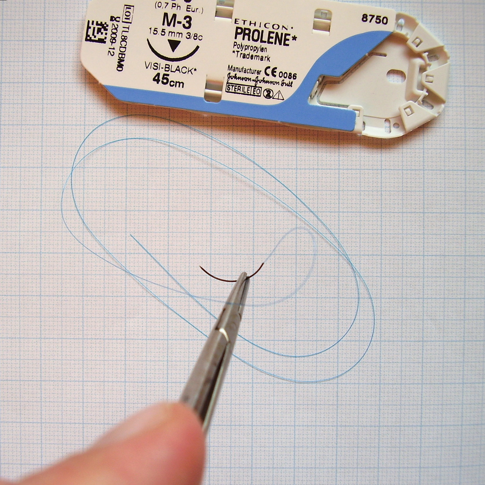
      suture

      From: Wikipedia - Surgical suture
      1. 清理傷口 要像小姐一樣大力...
      2. 蓋洞巾, irrigation, 三消. 看extend到哪 至少用摸的 照x光可看air, metal
      3. 打local
        之前看一本 臨床小秘笈 : 各科臨床技術的小撇步 還有長庚teaching 說打wound中央
        但 Clinical procedures on emergency medicine 跟學姊是說打進針點
        所以我就combine 目前少量樣本顯示 打wound中央+進針點 病人都不會痛XD 都打那麼多了...
        Clinical procedures on emergency medicine 還提到:
        grossly contamination 不要去碰wound, 但是一般L/W 會在"grossly" contamination還縫起來而嗎? 所以應該是都可以
        打進針點也有兩種方法:沒汙染的從wound邊緣的皮下進入;有汙染的(grossly)不要碰到wound 自wound 旁的皮 斜斜插入
        • local: 2% xylocaine 一般5cc以內。抽local的針跟打的不同,因為抽完針頭可能會頓掉。三消到一半可先打local,不一定要消完再打,病人不適會稍微少些
        • 小孩可能要先讓她睡著(sedation)
        • digital block: 手指nerve從兩旁走 所以兩邊手指跟部各打一次(1~2cc);而 youtube 上的影片是,從指背入,碰到骨頭收回1mm 再往左往右打。不管怎樣,注意不要打到血管,打得對皮膚會腫起來
        • 而 Emerg Med J 2010 Jul; 27:533. 提到了種新方法:再指頭最靠近手掌的指節的掌側,打2~3ml的local,但是這篇paper也提到他沒法收到預計的500人 且較遠的部份可能會麻不夠
        a single SC inj. of 2~3 mL of local anesthetic on the palmar surface at the base of the digit just distal to the proximal skin crease, followed by massage of the anesthetic into the area. because this method does not block the dorsal branches of the digital nerve, it might be less effective for injuries proximal to the distal-interphalangeal joint.
      4. suture (多出自長庚teaching)
        線:
        • 常用 3-0, 4-0.
        • 臉多用5-0, 6-0, 舌3-0,
        • oral mucosa用軟線.
        • scalp 年輕人3-0, 老人小孩4-0 (但馬偕都2-0) 線留長些.
        • 皮下之下用 dexon, vicryl (multifilament, absorbable 約30天)

        • scalp可用頭髮綁,之後再用tissue adhesive. indication: 符合下列條件:1)頭髮要夠長 2) 邊緣不可有挫傷 3) 無 grossly contamination 4) 已止血 5) 未傷到 galea (occipitofrontalis aponeurosis) 6) skull 沒有 fx
          但如果要打local確認wound下方組織,那還是suture起來的好(Clinical procedures on emergency medicine 及 Ann Emerg Med 2002; 40:19)

        持針: 在 Clinical procedures on emergency medicine 及 NEJM 2006;355;17 都建議用 open 拿法,食指去壓持針器的"關節" 用食指控制方向

        入針: 拿在距離線端1/2~1/3處. 針頭與皮膚成90度. 用forcep 去evert時,只能用一邊撐著 兩邊夾下去會傷害組織. 針到中央,再從中央拿針,用forcep evert另一側面 再繼續
        深度要大於寬度 底部要比表皮寬 間距=寬度

        傷口小其實不一定要用forcep, 這樣反而很卡
        一針到底~ 可用大拇指去壓對側皮膚 也可evert 但要小心被刺到
        出針後 用力拉起線的兩端 也可有 evert的效果 但也不要太緊@.@

        打結至少4個
        nylon, prolene易鬆脫 要多打幾個結

        剪刀: 一樣用食指壓 姆指向前 其他向後

      5. 拆線: 臉5~7天 身體10~14天 手掌>2wks, hip&knee 要很久

      2011年1月2日 星期日

      Shock

      EMPIRICAL CRITERIA FOR DIAGNOSIS OF CIRCULATORY SHOCK** Regardless of cause. Four criteria should be met.
      Ill appearance or altered mental status
      Heart rate >100 beats/min
      Respiratory rate >20 breaths/min or Paco2 <32>4 mM/L
      Urine output <0.5>20 minutes duration

      DEFINITIONS AND CRITERIA FOR SEPTIC, HEMORRHAGIC, AND CARDIOGENIC SHOCK
      • Septic Shock
        • Systemic Inflammatory Response Syndrome (SIRS)
        • 有兩個以上:
          1) Temperature >38℃ or <36℃>90 beats/min
          3) RR >20 breaths/min or Paco2 <32>12,000/mm3, <4000/mm3,>10% band neutrophilia
        • Severe Sepsis
        • SIRS + suspected or confirmed infection + associated with organ dysfunction or hypotension; organ dysfunction may include presence of lactic acidosis, oliguria, or altered mental status
        • Septic Shock
          SIRS + suspected or confirmed infection + hypotension despite adequate fluid resuscitation; septic shock should still be diagnosed if vasopressor therapy has normalized blood pressure
      • Hemorrhagic Shock
        • Simple Hemorrhage
          Suspected bleeding + pulse < 100
        • Hemorrhage with Hypoperfusion
          Suspected bleeding + base deficit <−4 mEq/L or persistent pulse >100 beats/min
        • Hemorrhagic Shock
          Suspected bleeding + Circulation Shock

      • Cardiogenic Shock
        • Cardiac Failure
          Clinical evidence of impaired forward flow of the heart, including presence of dyspnea, tachycardia, pulmonary edema, peripheral edema, or cyanosis
        • Cardiogenic Shock
          Cardiac failure + Circulation Shock


      CLINICAL MANAGEMENT GUIDELINES FOR FOUR COMMON CAUSES OF SHOCK
      • Hemorrhagic Shock
        1) Ensure adequate ventilation/oxygenation
        2) Provide immediate control of hemorrhage, when possible (e.g., traction for long bone fractures, direct pressure)
        3) Initiate judicious infusion of isotonic crystalloid solution (10–20 mL/kg)
        4) With evidence of poor organ perfusion and 30-minute anticipated delay to hemorrhage control, begin packed red blood cell (PRBC) infusion (5–10 mL/kg)
        5) With suspected central nervous system trauma or Glasgow Coma Scale score <9, immediate PRBC transfusion may be preferable as initial resuscitation fluid
        6) Treat coincident dysrhythmias (e.g., atrial fibrillation with synchronized cardioversion)

      • Cardiogenic Shock
        1) Ameliorate increased work of breathing; provide oxygen and positive end-expiratory pressure (PEEP) for pulmonary edema
        2) Begin vasopressor or inotropic support; norepinephine (0.5 μg/min) and dobutamine (5 μg/kg/min) are common empirical agents
        3) Seek to reverse the insult (e.g., initiate thrombolysis, arrange percutaneous transluminal angioplasty)
        4) Consider intra-aortic balloon pump counterpulsation for refractory shock

      • Septic Shock
        1) Ensure adequate oxygenation; remove work of breathing
        2) Administer 20 mL/kg of crystalloid or 5 mL/kg of colloid, and titrate infusion to adequate central venous pressure and urine output
        3) Begin antimicrobial therapy; attempt surgical drainage or d?bridement
        4) Begin PRBC infusion for hemoglobin < 8 g/dL
        5) If volume restoration fails to improve organ perfusion, begin vasopressor support; initial choice includes dopamine, infused at 5–15 μg/kg/min, or norepinephrine, infused at 0.5 μg/min


      Ref: Marx: Rosen's Emergency Medicine, 7th ed. Chp. 4 Shock